Sunday 27 April 2014

Elbow Fractures

Radiographs
 

1.    Anterior and posterior fat pads
 - If the anterior is elevated treat it as abnormal
 - If there is a posterior fat pad at all, treat as abnormal as 75% have fractures.

2.    Anterior humeral line
 On the lateral, draw a line down the anterior surface of the humerus. 1/3 of the capitellum should lie above the line.

3.    Radiocapitellar line
Draw a line through the centre of shaft of radius on either view. It should cut through capitellum on AP + lateral views.

4.    Ossification centres
As well as the bones, children have an added layer of complexity because of the ossification centres. The ossification centres tend to appear at these ages NOT fuse at these ages.

5.    Radial head, olecranon, distal humerus

Supracondylar Fractures: Ortho or # Clinic

Remember a displaced anterior fat pad, any posterior fat pad or an abnormal anterior humeral line are indications of an undisplaced supracondylar fracture (or a radial head fracture) and should be treated as such with either an above elbow POP or a broad arm sling if pain is not a significant feature.

Brachial artery injury in 5-10% Type II and Type III injuries. 

Radial (~25%) and ulnar (~15%) nerve damage unlikely. 60% of median nerve damaged. Mostly just motor branch, damaging intrinsic hand muscles so can't flex thumb IP joint.
In your clinical examination include:
-  a ‘pinch’ (medial nerve / AION),
-  ‘thumbs up’ (radial nerve), and
-  ‘give me four’ (finger abduction against resistance – ulnar nerve).
 
Complications include:
  • Cubitus varus (gun stock deformity)
  • Malunion and stiffness
  • Myositis ossificans
  • Nerve injury (most commonly median nerve)
  • Brachial artery (due to stretch and posterior displacement)
  • Volkmann’s ischaemic contracture (due to compartment swelling)
     
Olecranon Fractures: Ortho or #Clinic
Manage conservatively with POP. If significant displacement, discuss with orthopaedics.

Elbow dislocation: Ortho


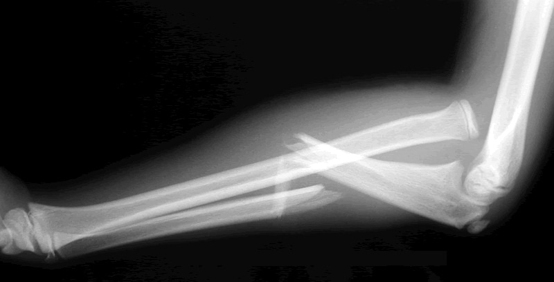
Elbow dislocations are surprisingly common, comprising 10 - 25% of all elbow injuries
The radial head may dislocate from the capitulum of the humerus on its own or in combination with dislocation of the ulna from the trochlea. The latter is usually straightforward to identify, but radial head dislocations may be more subtle - check the radiocapitellar line.

Radial Head: Fracture Clinic
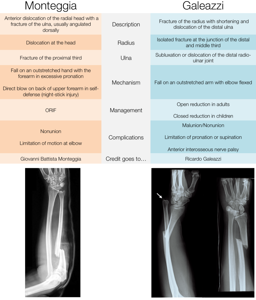
These injuries usually follow a fall onto an outstretched wrist or direct trauma. Radial head fractures usually occur in adults and account for 30% of all adult elbow fractures. It is the second most common elbow fracture in children. Examination may reveal local bruising and swelling. In some cases pain may only be evident with palpation of the radial head during passive forearm pronation. Elbow extension is usually restricted. Assessment of the wrist should be performed due to the possibility of an Essex-Lopresti fracture-dislocation, consisting of a comminuted radial head fracture with subluxation of the distal end of the ulna.

Undisplaced fractures can be managed with a collar and cuff sling and orthopaedic outpatient follow-up. Radial neck fractures: greater than 20° of angulation in the adult or 30° in the child requires reduction.

Medial epicondyle avulsion injury: Fracture Clinic
These injuries tend to occur in adolescents due to valgus stress during a fall on an outstretched hand. There may be associated ulna nerve damage and sometimes dislocation. Undisplaced avulsions can be managed conservatively while displaced fragments should be referred for reduction.

Pulled elbow: No follow up
The radial head dislocates from the annular ligament. In 50% of cases there is no history of a pull on the arm. The x-ray is normal and therefore not necessary if clinical suspicion is high prior to attempted manipulation.

There are 2 methods for reduction;
1. Supination of the forearm followed by flexion of the elbow
2. Hyperpronation of the wrist followed by flexion of the elbow - thought to be less painful. Elbow can start at 90, or be extended. Position one hand with the thumb over the radial head.


References
http://radiologymasterclass.co.uk/tutorials/musculoskeletal/x-ray_trauma_upper_limb/elbow_fracture_x-ray.html

http://www.enlightenme.org/knowledge-bank/cempaedia/elbow-injuries
http://www.enlightenme.org/learning-zone/pinch-me-thumbs-and-give-me-four
http://lifeinthefastlane.com/elbow-dislocation/
http://academiclifeinem.com/tricks-of-the-trade-nursemaid-elbow-reduction/
http://radiopaedia.org/articles/elbow-dislocation
http://journals.lww.com/em-news/blog/theproceduralpause/pages/post.aspx?PostID=8
http://emupdates.com/2009/09/19/emcard2054/
http://radiopaedia.org/articles/pulled-elbow-syndrome
http://emergencyeducation.net/1/post/2014/03/pulled-elbow.html
http://bestbets.org/bets/bet.php?id=123
http://pedemmorsels.com/nursemaids-elbow-feel-like-mr-miyagi/
http://www.ozemedicine.com/wiki/doku.php?id=elbow_pulled
http://academiclifeinem.com/tricks-of-the-trade-nursemaid-elbow-reduction/
http://radiopaedia.org/articles/supracondylar-fracture
http://emrems.com/tag/supracondylar/
http://lifeinthefastlane.com/bone-and-joint-bamboozler-007/
http://radiopaedia.org/articles/supracondylar-fracture-classification-1
http://radiopaedia.org/cases/supracondylar-process-of-humerus-1
http://radiopaedia.org/articles/olecranon-fractures-1
http://radiopaedia.org/cases/olecranon-fracture-2
http://radiopaedia.org/cases/olecranon-and-radial-head-fracture
http://radiopaedia.org/articles/radial-head-fractures

Tuesday 8 April 2014

Forearm Fractures

It's difficult to separate the radius from the ulnar as you progress up the forearm but isolated fractures can occur. Forearm fractures frequently seen in school aged children after a fall.  Adult fractures may be markedly displaced with little or no bony contact between the fragments. ORIF needed

Isolated ulnar fracture
Rare but can happen if the arm is in a defensive position, as in martial arts. POP. If displaced or angulated, for ORIF.

Isolated radial fracture
Very rare

Monteggia Fracture
    

Ulnar fracture and radial dislocation. You need to look carefully at the radiocapitella line. If it does not transect the middle of the capitlellum, there is a radial head dislocation. Occurs by forced pronation of the outstretched forarm. Urgent ORIF needed.


The Hume fracture is related - anterior dislocation of the radial head + olecranon fracture.

Galeazzi - radial fracture, dislocation at distal radioulnar joint. This occurs in teenagers and is very rare.

Galeazzi-v-Monteggia-XRays
http://www.pemcincinnati.com/blog/fracture-fridays-monteggiazzi/
Remember by:
" MUGR (Mugger): Monteggia Fracture = Ulnar fracture with radial head dislocation; Galeazzi Fracture = Radius fracture with DRUJ dislocation."


Monday 7 April 2014

The Radius


The radius is commonly fractured at the wrist. Because of the close connections with the ulna, breaks often happen together - at the wrist, the radius is the bone that is important, and is used to categorise the break. 

Colles Fracture
Radius within 2.5cm of the wrist. Distal fragment angulated to point dorsally.
Analgesia, POP, elevation
Discharge undisplaced

Complications: stiffness, malunion, reflex sympathetic dystrophy (Sudeck's atrophy) - refer for physiotheraphy, carpal tunnel syndrome, extensor pollicis longus rupture


Smith's Fracture
Unstable distal radius fracture (or reverse Colles' fracture)
The distal fragment is impacted, tilted anteriorly.
From a fall onto a flexed wrist.
Analgesia, POP, orthopaedics


Barton's Fracture
Intra-articular fracture involving the volar portion of the distal radius. The resultant fragment slips anteriorly, so the fracture is unstable.
POP and refer 


The Carpal Bones


http://www.instantanatomy.net/arm/areas/hand/carpalbones.html

Fractures are 10 times less likely to occur than distal radial and ulnar injuries and are rare in children under 12. Injuries more likely in the proximal carpal row (scaphoid, lunate, triqetrum, pisiform).

TriquetrumThis is the second most fractured carpal bone accounting for 20%Z of carpal injuries. May be related to forced hyperextension. Normally seen as small triangular fragements or flake avulsions.
Treat with immobilisation in a POP backslab or wrist support splint, analgesia and refer to fracture clinic.

HamateThis is often caused by axial loading or punch type injury. Suspect if there is local palmar tenderness. Diagnosis can be difficult.

LunateFractures are uncommon, and mechanism is typically a direct blow to the carpus. Avascular necrosis may occur.

Pisiform
Direct blow or FOOSH.

Capitate

Axial loading of the middle metacarpal. Often missed.

Trapezoid
Rare. Axial loading of the index metacarpal.

Trapezium
Rare. Forced abduction of the thumb. Can mimic a scaphoid fracture.

All of these injuries should be referred to plastics?

http://www.memrise.com/course/59292/anatomy-of-the-hand/

Olecranon Fractures

The Olecranon is the proximal part of the humerus. It is rare to fracture it. Olecranon fractures usually occur after
- direct blow or fall onto the elbow
- fall on outstretched hand with elbow flexed
- avulsion fracture
- stress fracture eg. throwing, weight lifting

Clinical Features
Significant amounts of elbow swelling
Inability to extend arm (due to triceps rupture)
Ulnar nerve injury possible


Radiological Findings
Fracture is normally obvious.

The radiological difficulty is differentiating fractures from the olecranon epiphysis. The olecranon epiphysis appears between the age of 8 and 11, and fuses by the age of 14.


Management
Undisplaced needs above elbow backslab, and fracture clinic.
Displaced or involving then elbow joint need an ORIF - so immediate orthopaedic opinion.

References

http://radiopaedia.org/articles/olecranon-fractures-1
http://radiopaedia.org/cases/olecranon-fracture-2
Oxford Handbook of Emergency Medicine
Minor Injuries
Musculoskeletal Trauma
Emergency Care of Minor Trauma in Children


Sunday 6 April 2014

Scaphoid Fractures

There's loads of really good FOAMEd resources out there already about scaphoid fractures. It's common (2-7% of all fractures), and most commonly occurs in young people as result of FOOSH.

Anatomy - look at Emergency Medicine Ireland. It's fabulous, why do anything else?
 
Clinical Examination
Clinical examination is rubbish, but a combination of ASB tenderness (90% sensitivity, 40% specificity), thumb axial compression and scaphoid tubercle tenderness (57% specificity) is the best way.

Imaging
You might see:
- visualisation of the fracture +/- displacement
- soft tissue swelling and lateral displacement of the adjacent fat pads
- scaphoid fat pad sign - formation of a straight/convex line adjacent to the concave aspect of the scaphoid
- associated scapholunate ligament disruption (Terry Thomas sign)
Helpfully, 25-30% have normal x-rays.
Ultrasound is not sensitive enough. Most places go for CT  or MRI.

Management
If radiologically confirmed, no displacement, POP and fracture clinic.
        Distal 1/3 -  6-8 weeks.
        Middle 1/3 – 8-12 weeks.
        Proximal 1/3 – 12-23 weeks.
If radiologically confirmed + displacement, speak to plastics.

If no radiological evidence, splint and bring back to fracture clinic. Thumb extension is not needed, as long as there is no ulnar deviation.

Complications
Avascular necrosis occurs in 30-40% of untreated fractures. It is less than 10% in undisplaced fractures. Non-union occurs in 5-12% of treated fractures, and the incidence of complications increases with proximal pole fractures.
80% of patients are immobilised unnecessarily.

References
http://www.enlightenme.org/knowledge-bank/cem-ctr/early-ct-clinically-suspected-scaphoid-fractures
http://bestbets.org/bets/bet.php?id=2003
http://bestbets.org/bets/bet.php?id=1687
http://bestbets.org/bets/bet.php?id=1673
http://lifeinthefastlane.com/scaphoid-fractures-the-ed-perspective/
http://emergencymedicineireland.com/2012/06/anatomy-for-emergency-medicine-19-scaphoid-fractures/
http://blogs.bmj.com/bjsm/2013/12/16/current-management-of-occult-scaphoid-fractures-in-uk-emergency-departments/
http://radiopaedia.org/articles/scaphoid_fracture
http://foam4gp.com/2013/11/15/foam4gp-map-scaphoid-fractures-thumbing-through-the-research/
http://radiologymasterclass.co.uk/tutorials/musculoskeletal/x-ray_trauma_upper_limb/wrist_trauma_x-ray.html
http://shortcoatsinem.blogspot.co.uk/2012/03/heeling-scaphoid-terrible-pun-intended.html

Abdominal and Orbital Compartment Syndrome

Abdominal compartment syndrome often follows laparotomy. Tissue swelling combined with packing or haematomas may cause rising abdominal pressures, causing cardiovascular, respiratory, renal and cerebral dysfunction.
It is best noticed by a fall in urine output + raised JVP

Increased Risk If:
Diminished Abdominal Wall Compliance

    acute respiratory failure (especially with elevated intrathoracic pressure)
    abdominal surgery with fascial closure
    major trauma/burns
    prone positioning
    head > 30 degrees
    high BMI
    central obesity
Increased Intra-luminal Contents

    gastroparesis
    ileus
    colonic pseudo-obstruction
Increased Abdominal Contents

    haemo/pneumoperitoneum
    ascites
    liver dysfunction

Capillary Leak or Fluid Resuscitation

    acidosis (pH < 7.2)
    hypotension
    hypothermia (T < 33 C)
    massive transfusion (> 10 U in 24 hrs)
    coagulopathy
    massive fluid resuscitation
    pancreatitis
    sepsis
    oliguria
    damage control laparotomy


Orbital Compartment Syndrome


Retrobulbar hemorrhage with acute orbital compartment syndrome is primarily a clinical diagnosis. CT may show a diffuse, increased reticular pattern of the intraconal orbital fat rather than a discrete hematoma.
A teardrop or tenting sign is ominous - it occurs when the optic nerve is at maximum stretch and distorts the back of the globe into a teardrop shape.

The main steps in emergency canthotomy/ cantholysis are:
        use local anesthetic but warn the patient that they may feel pain
        Perform the canthotomy:
            place the scissors across the lateral canthus and incise the canthus full thickness
        Perform cantholysis:
            Grasp the lateral lower eyelid with toothed forceps
            Pull the lower eyelid anteriorly
            Point the scissors toward the patient’s nose, place the blades either side of the lateral canthal tendon, and cut
http://ccforum.com/content/4/1/23
http://lifeinthefastlane.com/ophthalmology-befuddler-033-2/

Compartment Syndrome

Compartment syndrome is without doubt one of the diagnoses that we should never miss. Palpable distal pulses and normal capillary refill times do not exclude compartment syndrome.

Pathophysiology
The pressure inside a body compartment increases. The compartment can't compensate for the increase in pressure, so puts pressure on nerves and blood vessels, making limbs painful and ischaemic. 

Compartment syndrome can affect many regions of the body and is more common in:
- the forearm (volar compartment): humeral shaft fracture, radius and ulna fracture, supracondylar fracture
- leg (the anterior compartment): tibia fracture
It can occur in other regions, including the back, buttocks, thigh, abdomen and foot. Compartment syndrome of the deep posterior compartment of the leg is easily missed.

Compartment syndrome can also occur after soft tissue injuries:
    crush injury
    snake bite
    excessive exertion and exercise - used to be called anterior tibial syndrome. Pain persists despite rest.
    prolonged immobilisation
    constrictive dressings and plaster casts
    soft tissue infection
    seizures
    extravasation of intravenous fluids and medications
    burns
    tourniquets
Patients with a coagulopathy are at particular risk of compartment syndrome.

DiagnosisSuspicion is essential! Suspect if:
 -       one of the fractures listed above is present
 -       one of the soft tissue injuries listed above is present (e.g. crush injury)
 -       patient has a coexistent coagulopathy

Examination - The 6Ps    1. pain - Occurs early, is persistent, tends to be disproportionate compared with the original injury and is not relieved by immobilisation. Passive stretching worsens the pain.
    2. pallor
    3. perishingly cold
    4. pulselessness (a late sign)
    5. paralysis
    6. paraesthesiae - assess by light touch and two-point discrimination, rather than just pinprick, which is less sensitive.
The extremity may be swollen and affected compartments may feel tense and tender on palpation.
  
Pressures:
zero - 10 mmHg = normal tissue pressure
>20mmHg = capillary blood flow may be compromised
>30 - 40mmgHg = muscle and nerve fibers at risk for ischemic necrosis at pressures

Thinking about the Whiteside' Theory:
"the development of a compartment syndrome depends not only on intra-compartment pressure but also depends on systemic blood pressure" treatment may depend on calculating the delta pressure.

Delta pressure = diastolic blood pressure (DBP) — intracompartment pressure
A delta pressure <30 mmHg is suggestive of compartment syndrome

Treatment
This is a surgical / orthopaedic emergency - ischemic injury to muscles and nerves occurs after 4 hours of complete ischemia and becomes irreversible over the next 4 hours, resulting in local rhabdomyolysis and neuropraxis.

Remove all constrictive dressings
Elevation
Traction as appropriate
Analgesia
IV hydration to relieve rhabdomyolysis


References

http://lifeinthefastlane.com/ortho-library/compartment-syndrome/
http://radiopaedia.org/articles/chronic-exertional-compartment-syndrome
http://www.bmj.com/content/325/7364/557?sso=
http://www.trauma.org/archive/resus/DCSacs.html
http://lifeinthefastlane.com/bone-and-joint-bamboozler-002/
http://lifeinthefastlane.com/education/ccc/abdominal-compartment-syndrome/
http://journals.lww.com/anesthesia-analgesia/Fulltext/1996/12000/Acute_Biceps_Compartment_Syndrome_Associated_with.40.aspx
http://bjsm.bmj.com/content/38/2/218.full
http://pedemmorsels.com/compartment-syndrome/
http://calgaryguide.ucalgary.ca/slide.aspx?slide=Acute%20Compartment%20Syndrome.jpg